Since the 1930s, the so-called homunculus map has shown how different parts of the brain’s motor cortex may control movement to different parts of the body. But it may be missing an important network
By Jason Arunn Murugesu
19 April 2023
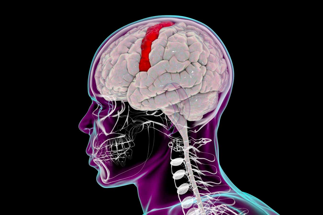
Human brain with a highlighted precentral gyrus, the location of the motor cortex
Science Photo Library/Getty Images
Our movements may be controlled by two distinct networks in our brain, rather than just one.
For nearly a century, we have known that the motor cortex – a thin strip of tissue that runs across the top of the brain – controls our body movements.
In the 1930s, neuroscientists Wilder Penfield and Edwin Boldrey electrically stimulated the brains of people undergoing brain surgery, showing that different parts of the primary motor cortex control different parts of the body. They also found that these control areas are arranged in the same order as the body parts they direct, with the toes at one end and the face at the other, as depicted by the so-called homunculus map, or a likeness of a person, they drew to represent this pattern (see image below).
Advertisement
Evan Gordon at Washington University School of Medicine in Missouri and his colleagues wanted to use modern technology to look into the Penfield-Boldrey idea in more detail. They therefore took high-resolution MRI brain scans from seven people as they lay in a scanner looking at a cross symbol for a total of 12 to 15 hours over multiple sessions.
Read more:
Roots of five medical conditions shown in map of the developing brain
Analysing the participants’ brains while they were largely stationary and not engaged in a more sophisticated task, such as reading, meant that their brain activity data was less complex, allowing the researchers to better observe which parts of the brain work together, says Gordon.